SonoPhile
a 52-year-old man admitted with severe abdominal pain and an unremarkable medical history. Initial ultrasound, CT, MRI, and lab tests suggested a paraduodenal abscess or gastrointestinal stromal tumor. Referred for endoscopic ultrasonography, a retroperitoneal foreign body—a toothpick—was discovered and removed.
Unnoticed foreign body ingestion


Transabdominal ultrasound, Aloka Alpha Prosound 6, straight arrow: stomach (pyloric part) transverse view, curved arrow: toothpick
Initial Hospitalization:
-
Symptoms: Strong abdominal pain.
-
Lab Results: High serum CRP (193 mg/l) and elevated WBC count; normal pancreatic enzyme activity, bilirubin, AST, ALT, and GGTP levels.
-
Ultrasound: Hypoechoic, polycyclic lesion (35 × 28 × 50 mm) near the pancreatic body and tail.
-
CT Scan: Large pathological mass (50 × 40 × 50 mm) near duodenum, superior mesenteric artery, pancreatic body; possible paraduodenal abscess or GIST.
-
MRI: Homogenous contrast enhancement, low ADC map signal, restricted diffusion, more consistent with GIST than abscess.
-
Treatment: Antibiotics, resulting in decreased serum CRP levels and reduced pain.
Follow-up Admission:
-
Delay: Patient delayed EUS-related hospitalization for two weeks, showing up asymptomatic with normal lab results on the day of admission.
-
Transabdominal US: Linear hyperechoic object (40 mm) from gastric antrum wall towards pancreatic body, surrounded by dense fluid collection, indicating a foreign body.
-
Medical History: Patient initially could not recall ingesting a foreign body.
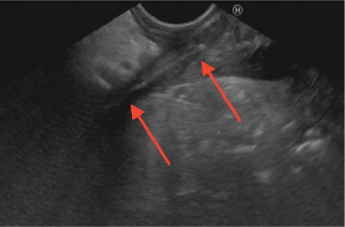
Endoscopic ultrasound, Hitachi Avius, Pentax EG-3870 UTK, transgastric approach, arrows: toothpick
Credit to: Tynecki, W., Tynecki, A., Grobelna, A., Baranowski, T., Siemiaszko, G., Łapiński, T., & Wilczek, J. (2020). A case of ultrasound diagnosis of retroperitoneal lesion caused by unnoticed foreign body ingestion. Journal of ultrasonography, 20(81), e151–e153. https://doi.org/10.15557/JoU.2020.0024

-
Further Diagnostic Steps:
-
Gastroscopy: Showed no obvious abnormalities.
-
EUS Ultrasound: Confirmed linear foreign body stuck (5.6 mm deep) in the muscular layer of the antrum.
-
Detailed Evaluation: Lesion and surrounding structures were described.
-
-
Surgical Intervention:
-
Consultation: Patient eligible for laparotomy.
-
Procedure: Wooden toothpick (40 mm) extracted.
-
-
Rare Case: Unnoticed ingestion of a foreign body caused alimentary tract perforation with total displacement of the object out of the tract lumen.
-
Diagnostic Importance of Sonography: Sonographic evaluation is crucial in the diagnostic process, even after using cross-sectional contrast-enhanced imaging techniques.
-
Reevaluation Benefits: Repeating transabdominal ultrasound and adding endosonography helped in making the correct diagnosis.