SonoPhile

Post-operative Abdominal Pain
The case presented here describes a 9-year-old female who presented with fever and worsening abdominal pain 4 days after laparoscopic resection of a benign ovarian teratoma. Computed tomography failed to provide adequate diagnostic imaging. Ultrasound was subsequently used to rule-out a major post-operative complication and ultimately led to a successful non-operative approach while avoiding repeat radiation exposure.
Post-Operative Day 4:
-
Developed intermittent fevers (max 102.8°F), transient abdominal and shoulder pain
-
Imaging: Small-volume pneumoperitoneum, deemed normal; benign abdominal exam
-
Management: Watchful waiting at home with close follow-up
Post-Operative Day 5:
-
Symptoms: Persistent shoulder and abdominal pain, overnight fever (100.9°F), loose green stools
-
Hospital admission for observation
-
Imaging: CT abdomen/pelvis showed obscured lower pelvic mass, no contrast extravasation; poor sensitivity due to thin body habitus
-
Lab findings: Negative stool panel, normocytic anemia (10.1 g/dL, pre-op 11.3 g/dL)
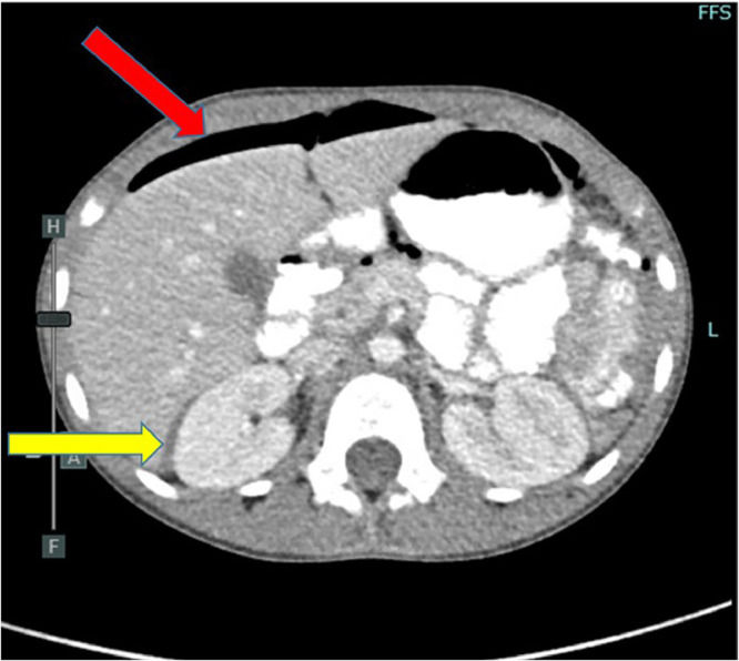

Axial view – CT abdomen/pelvis with IV and PO contrast demonstrating small-volume pneumoperitoneum (red arrow) and complex free fluid in the hepatorenal space (yellow arrow).
Coronal view – CT abdomen/pelvis with IV and PO contrast demonstrating complex hyperdense mass external to bowel located within the pelvis (yellow arrow).
Post-Operative Day 6 (Hospital Day 2):
-
Acute worsening of abdominal pain with rebound tenderness
-
Imaging: Abdominal and transabdominal pelvic ultrasound revealed:
-
Complex free fluid in hepatorenal space
-
Focal hematoma in lower pelvis (7.5×6.6×4.3 cm) without internal Doppler flow, suggesting low likelihood of active bleed
-
-
Discharged home with close follow-up, no further post-operative issues

Right upper quadrant transabdominal ultrasound demonstrating complex free fluid in the hepatorenal space 1.9-cm wide suggesting hemoperitoneum.

Right upper quadrant sagittal ultrasound demonstrating hemoperitoneum along the inferior hepatic margin.
Right lower quadrant sagittal ultrasound demonstrating heterogeneous mass external to bowel measuring 7.5 × 6.6 × 4.3 cm consistent with focal hematoma.

Right lower quadrant sagittal ultrasound demonstrating an absence of internal Doppler flow suggesting hematoma without active bleed.
Ultrasound vs. CT in Pediatric Post-Operative Care:
-
Ultrasound effectively elucidated abdominal and pelvic findings after CT scan ambiguity.
-
Role in triage: Determines need for operative intervention while minimizing radiation exposure.
-
Preference for contrast-enhanced ultrasound due to comparable accuracy to CT in detecting intraabdominal organ injury.
-
Ultrasound advantages: Provides detailed images of internal composition, vascularity, and relationships with adjacent structures.
-
Case example: Post-operative symptoms attributed to peritoneal irritation by hematoma identified via ultrasound; successful non-operative management.
Conclusion
Credit to: White, A. B., Bacon, D. R., Olinger, K., & Dehmer, J. J. (2022). A case report on ultrasound evaluation of pediatric post-operative abdominal pain. Radiology case reports, 17(11), 4223–4226. https://doi.org/10.1016/j.radcr.2022.08.040