SonoPhile

Single Accessory Splenic Infarction in a Patient with Accessory Spleens in the Abdominal Cavity
A 19-year-old male with infarction in one of four accessory spleens. Imaging diagnosis was challenging, and definitive diagnosis came from postoperative pathology showing no torsion. After surgery and anti-inflammatory treatment, the patient recovered without complications at the 3-month follow-up. This case underscores the difficulty in diagnosing accessory splenic infarction without torsion using imaging alone, suggesting a multimodality approach and diffusion-weighted imaging for diagnosis confirmation.
-
Initial Presentation:
-
Symptoms: Sharp left upper abdominal pain after heavy eating and drinking, lasting 3 days.
-
Initial Treatment: Anti-inflammatory medication and analgesia relieved pain.
-
-
Initial Diagnostic Imaging:
-
CT Scan (Day 3): Mass (4.5 cm × 3.0 cm × 6.7 cm) with well-defined margin, homogeneous soft tissue density, and edematous surrounding fat tissue.
-
Enhanced CT Scan (Day 6): Increased mass size (4.8 cm × 3.3 cm × 7.4 cm) with pronounced annular enhancement; found three accessory spleens.
-
-
Admission to Hospital:
-
Timing: 14 days after initial episode.
-
Medical History: No operative treatment, trauma, family history of atrial fibrillation or coagulopathy.
-
Physical Exam: No tenderness, mass, or palpable liver; normal vital signs.
-
Lab Tests: Normal white blood cell count, hemoglobin, platelet count, liver enzymes, coagulation profile, D-dimer, and CRP levels; tumor markers within normal range.
-

Conclusion
-
Condition: Accessory splenic infarction without pedicle torsion is extremely rare.
-
Imaging Complexity: Imaging characteristics vary with different stages of infarction.
-
Diagnostic Tools: Ultrasound, CT, and MRI provide valuable insights
Credit to: Xu, N., Xu, Y., & Zhu, Q. (2023). Radiologic Findings of Single Accessory Splenic Infarction in a Patient with Accessory Spleens in the Abdominal Cavity: A Case Report. Medicina (Kaunas, Lithuania), 59(4), 807. https://doi.org/10.3390/medicina59040807
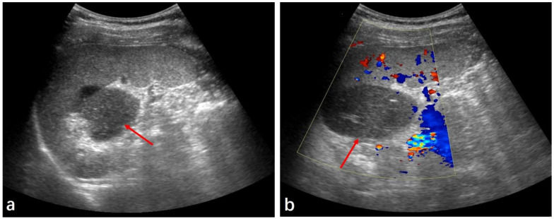
(a) Ultrasound showed a solid mass at the splenic hilum (red arrow). (b) Color Doppler showed no significant blood flow (red arrow).
Further Imaging:
-
Abdominal Sonography (Day 12): Large, well-defined solid mass with low inhomogeneous echogenicity, no blood flow observed.
-
MRI (Day 12): Hypointense on T1WI, hyperintense on T2WI, scattered hyperintense strip indicating hemorrhage, size reduced slightly (4.6 cm × 3.1 cm × 7.0 cm), high degree of diffusion limitation, no abnormalities in other organs or lymph nodes.
Diagnosis and Treatment:
-
Surgical Findings: Mass fed by single vascular pedicle connected with splenic artery, non-twisted pedicle, mass with complete capsule and gray-red surface.
-
Microscopic Findings: Large patches of infarcted areas, peripheral fibrous proliferation, slight vasculature.