SonoPhile
Bladder

To assess
Assess the urinary bladder for wall hypertrophy, intraluminal findings, and urinary retention (as applicable).
Limitations
None
Patient's Preparation
• Patient should be well hydrated.
• The patient should be kept from voiding 30 minutes before the study.
• If the patient has a suprapubic or Foley catheter, the catheter should be clamped 1 hour before the exam to allow for bladder filling unless otherwise contra-indicated based on provider instructions/preferences.
Equipment Setup
Curvilinear transducer with a frequency range of 2-9 MHz that allows for appropriate penetration and resolution of anatomy, depending on patient’s body habitus
Common Pathology
-
Postrenal acute kidney injury
-
Voiding dysfunction
-
Estimation of bladder volume
-
Haematuria
-
Confirmation of proper urinary Foley catheter placement
-
Malfunctioning Foley catheter
Scanning Technique
-
Longitudinal View
-
Place the transducer with the indicator pointing towards the patient’s head in the patient’s midline, right above the pubic symphysis.
-
Rock the probe so that it points down towards the pelvic cavity.
-
Observe the lateral borders of the bladder by tilting the probe left and right.

2. Transverse View
-
Next, centre the bladder and then rotate the transducer 90 degrees counterclockwise. The indicator should now point to the patient’s Right side.
-
Make sure to tilt the ultrasound probe so it scans into the pelvic cavity.
-
Tilt/Fan the probe to examine the entire bladder from superior to inferior.

3. Calculate the Volume of the Bladder
-
Most ultrasound machines will automatically calculate the entire bladder volume, which should be less than 300-400 mL in healthy adults, and Post Void Residual (PVR) should be less than 50-100mL (Fitzgerald; Latini).

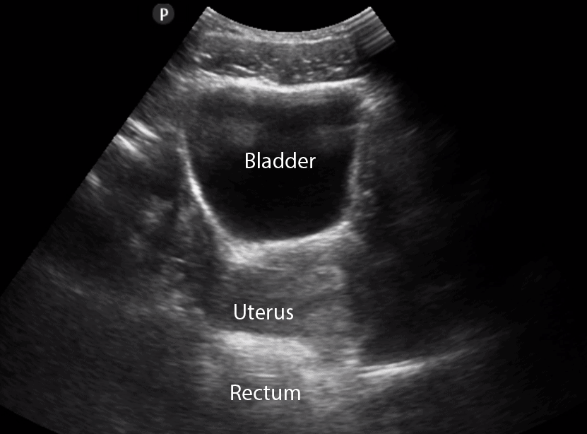
Longitudinal view: Female Bladder
Longitudinal view: Male Bladder

Transverse view: Male Bladder
Transverse view: Female Bladder